The Composition Of The Hippocampus: Structure And Functions
Cognitive processes such as learning and remembering are crucial for human beings. For example, the hippocampus plays a fundamental role in these processes. This is one of the areas in the composition of the hippocampus.
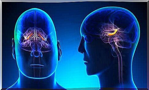
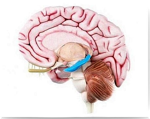
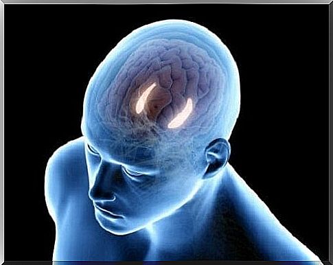
The composition of the hippocampus is a distinctive C-shaped structure. It is located in the lower part of the lateral ventricle of the brain. The hippocampus itself consists of three main parts (CA1 – CA3). Does hippocampal composition include only the hippocampus?
Anatomical analysis of the composition of the hippocampus
In the 16th century, the anatomist Arantius first spoke about the composition of the hippocampus. He also gave it the name hippocampus. This word comes from the Greek word for seahorses.
However, the composition of the hippocampus is not just the hippocampus. Actually, it also consists of the dentate gyrus, the subiculum and the entorhinal cortex.
The whole of the hippocampus is thus about five centimeters long. In the middle part is also the uncus, which is shaped like a potato. The uncus varies from brain to brain.
The formation of the hippocampus: the architecture
The dentate gyrus
The dentate gyrus is located most in the center of the cerebral cortex. In terms of cell formation, the dentate gyrus is a trilaminate brain region. In the hippocampus, the dentate gyry with its typical C-shape on the hippocampal fissure is centrally separated from the first part of the hippocampus and next to it from the subiculum.
The main cell layer of this structure is full of granular cell bodies. The apical dentrites of these cells have branches in the molecular layer of the dentate gyrus. The granular cells and the molecular layers together form the fascia dentata.
The third layer closest to the inside of the dentate gyry is the polymorphic layer or hilus. In addition, there is a part of the pyramidal cell layer that is enclosed by the granular cells.
The hippocampus
The parts of the hippocampus are called CA1, CA2, and CA3. They consist of one cell layer: the pyramidal cell layer. The surface is called the alveus. This is adjacent to the ventricular lumen formed by axons of pyramidal cells. Historically, this area consists of:
- Lucidum stratum
- Radiatum stratum
- lacunosum-moleculare
The lucidum stratum or CA3 has fibers that form nearby dentritic synapses above the pyramidal cell wall of this layer. The CA2 layer is fairly compact. This layer also shows a pyramidal cell layer but it is difficult to determine its borders.
Furthermore, the CA1 layer is also a part of the hippocampus. The pyramidal cell layer in this region, in turn, consists of one inner and one outer layer.
the subiculum
The CA1 layer and the subiculum overlap at the edges. In this way they form a transition zone. The subiculum is primarily divided into the following layers:
- On the surface there is a spacious molecular layer. This is where the dentrites of the pyramidal cells are located. This pyramidal cell layer can then be split into two underlying layers: an outer and an inner layer.
- The cells of the outer layer have an accumulation of lipofuscin pigment in their apical dentrites.
- The presubiculum consists of a superficial layer, which contains modified pyramidal neurons.
- The parasubiculum has one cell layer that is difficult to distinguish from the presubiculum.
The entorhinal cerebral cortex
The term “entorinal” is synonymous with Brodmann’s area. It extends mainly rostrally (on the side of the nose) towards the middle part of the amygdala. At the posterior, however, it extends towards the posterior commissure of the nucleus lateralis geniculatus. This area is slightly different from the rest of the brain areas.
The connection in the composition of the hippocampus
The intrinsic pathway of the hippocampus
The connection in the hippocampus follows a one-way and glutamatergic (outside) pathway that is part of a closed circuit. In this intrinsic chain of connections, first and foremost the dentate gyrus is very important. It receives the most information. This is then transmitted to the entorhinal cerebral cortex.
External connections
The extrinsic pathway of the hippocampus is formed by:
- Multiple cerebral arteries.
- The amygdala.
- The medial septal nucleus.
- thalamus.
- The supramammillary nucleus.
- The monoaminergic nucleus of the brainstem.
That’s apparently how the hippocampus receives sensory information from a variety of brain regions.
Cortical Connections
These projections serve first and foremost to bring the sensory information into the hippocampus.
Subcortical Connections
The fimbriae and fornix form the classical efferent system of the hippocampus. In addition, there are also enormous connections between the hippocampus and the amygdala. Furthermore, connections are also established along the subiculum between the hippocampus and the hypothalamus.
As you can see, the composition of the hippocampus is a complex set of regions that also include the hypothalamus. However, most research projects have studied animals. Still, it seems clear that the areas described here are very similar to the hippocampus in humans.