Biopsychological Research Methods
Biopsychological research methods have changed a lot in recent decades. Although several biopsychological research methods exist, in this article we focus on the methods that study what happens in the brain under certain conditions.
Authors such as Dewsbury (1991) define biopsychology as “the scientific study of the nature of behavior,” some refer to as psychobiology.
However, other authors prefer the term biopsychology because they say it “indicates a biological approach to psychology rather than a psychological approach to biology.”
Human brain stimulation and visualization methods
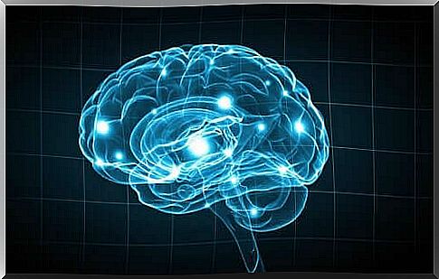
Being able to observe and record brain activity is a very important milestone that was achieved thanks to the various techniques that scientists developed in the twentieth century. These biopsychological research methods are without a doubt a major breakthrough in the study of our most curious organ.
Contrast radiography
This technique consists of injecting a substance into the body to absorb X-rays. In this way, scientists can see more clearly the contrast between this area and the tissue around it.
Cerebral angiography is a type of contrast radiography. To do this, a contrast agent is placed in a brain vessel. The goal is to observe the vascular system while performing an X-ray. This technique is very useful to locate vascular wounds and brain tumors.
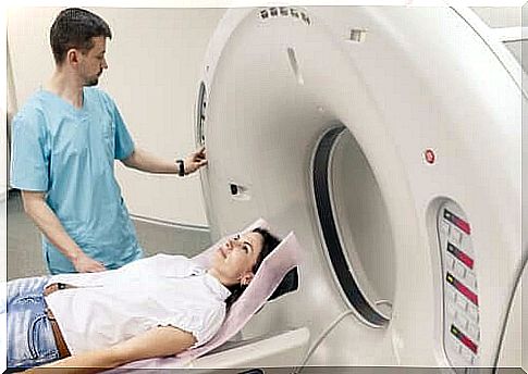
Computed tomotgraphy (CT scan)
Through a CT scan, experts can see the entire brain structure. For this examination, it is the intention that the patient lies down in the middle of a large tube.
While the patient remains as still as possible, several measurements are made by means of an X-ray source and an X-ray detector. This is done as the beam of radiation and the detector revolve around the patient’s head.
Then all the information is passed to a computer, allowing the doctors to examine the brain on a horizontal plane. Usually they do this on eight to nine horizontal brain sections. Once all the explorations have been combined together, it is possible to create a three-dimensional representation of the brain.
Nuclear Magnetic Resonance (NMR)
Thanks to the different waves that hydrogen atoms give off when they are activated by radio frequency on a magnetic field, NMR makes it easier to get a sharper image of the brain. It offers a high spatial resolution and produces three-dimensional images.
Positron Emission Tomography (PET)
PET allows us to obtain images of brain activity rather than brain structure. To obtain these images, scientists inject a radioactive isotope into the patient’s carotid artery. Active neurons quickly absorb this isotope.
Once the neurons stop metabolizing it, it accumulates and then slowly breaks down. In this way, you can observe which neurons are active at any given time while performing different activities.
Functional MRI
Using MRIs, on the other hand, we can get a picture of the increase in the amount of oxygen in the blood of the brain. MRIs are thus able to measure brain activity. When we compare it to PET, MRI actually offers four advantages:
- Doctors do not have to administer anything to the patient.
- It provides both functional and structural information.
- It offers better spatial resolution.
- MRI provides three-dimensional images of the entire brain.
Magnetoencephalography (MEG)
MEG measures the changes in the magnetic fields on the surface of the scalp. These changes occur as a result of the variations in the guidelines for neuronal activity.
Transcranial Magnetic Stimulation (TMS)
Walsh and Rothwell (2000) state that TMS “changes an area of the cortex, creating a magnetic field beneath the spiral that runs across the skull.” TMS temporarily ‘switches off’ a part of the brain, in order to study behavior and cognition under these conditions.
Lesion Methods
Lesion methods focus on destroying a small part of the brain to see how it affects behavior.
- Aspiration lesion. This method creates a lesion in an exposed or easily accessible area of cortical tissue. The doctors remove the tissue with a pointed pipette.
- Radiofrequency lesion. For this method, small subcortical lesions are created. To do this, an electrode directs the high-frequency current through the tissue to be examined. The size and shape of the lesion depend on three factors:
- Duration of the procedure.
- Intensity of the current.
- Configuration of the electrode tip.
- Skull cuts. In this case, the brain region in question is divided into several sections.
- Cooling lesions. Although this biopsychological research method falls under lesion methods, it is in fact temporary and reversible. Instead of destroying tissue , a brain region is cooled to just above freezing point. This causes the neurons to stop sending signals, leaving the cold area of the brain blocked. In this way, researchers can see which behavioral changes this brain region causes. Once the temperature returns to normal, brain function is restored.
Electrical stimulation
Another interesting biopsychological research method is electrical stimulation. This procedure consists of electrically stimulating a part of the nervous system to obtain data about its functions. Usually a bipolar electrode is used here.
This stimulation ‘shoots’ neurons and changes their behavior. In general, this method produces the opposite effect of lesion methods. For example, if it is possible to drastically reduce sleeping hours with a lesion, electrical stimulation sleeping behavior can become clumsy and more difficult to control.
Lesion methods with electrical recording
- Intracellular Uptake. In this technique, doctors place a microelectrode in the inner part of a neuron. It records fluctuations in the membrane potential.
- Registration of extracellular units. In this case, a microelectrode is placed in the extracellular fluid surrounding the neuron. However, it does not provide information about the membrane potential.
- Multi-unit recording. In this case, the electrode tip is larger than that of a microelectrode. This allows it to simultaneously pick up the signals from multiple neurons. The detected potentials are then put on a circuit where they are integrated.
- Invasive EEG Monitoring. Here stainless steel electrodes are inserted into the skull. For subcortical signals, the electrodes usually consist of cable. Doctors implant them through stereotactic surgery.
Biopsychological research methods: still a long way to go
In this article we have discussed some of the most important biopsychological research methods. However, it should be noted that there are also other biopsychological research methods that study other areas of the body, such as muscle tone, eye movement, skin conductance or cardiovascular activity.
The breakthrough in this area is undoubtedly spectacular, but not yet decisive. Perhaps in a few years scientists will come up with new techniques that contribute to the evolution of neuroscience, which will improve the quality of life of many people who are victims of brain injuries.